Skeletal muscles form about 40–45% of the total human body mass and are essential for sustaining life. Proper muscle function; allows mobility, joint stability, postural control, breathing, metabolic control, thermoregulation, and energy storage [1]. The incidence of traumatic skeletal muscle injuries is increasing dramatically throughout the world, and there may be many reasons for this. Sports-related injuries which are seen after surgical interventions such as tumor ablation, soft tissue reconstruction, or joint arthroplasty can be cited as examples [2–4]. Despite its high incidence, treating injured muscles and restoring their original structures and functions have not yet been fully achieved [5].
There are various conservative treatment strategies for acute and chronic skeletal muscle injuries, such as POLICE (Protection, Optimal Loading, Ice, Compression, and Elevation) [6], physiotherapy applications (exercise, stretching, mobilization, massage, ultrasound, laser, electrical stimulations, etc.), non-steroidal anti- anti-inflammatory drugs, biological repair agents and surgical methods [7, 8]. Physiotherapy methods are among the first preferred treatment options for muscle injuries because of being affordable, noninvasive, safe, and effective and have been used for many years.
Stretching exercises can be used for treatment as they can show positive effects at the cellular level with the intermittent tension applied during the acute recovery period. Although the benefits of stretching exercises on injured fiber are still unveiled, it has been proven in a limited number of studies that it stimulates collagen synthesis, which has an important role in healing [6].
In addition to the physiotherapy modalities commonly used in rehabilitation clinics, alternative treatment modalities are also being tried. Pulsed electromagnetic field (PEMF) therapy, one of the physiotherapy methods, has been used for a long time for avascular necrosis, fracture, soft tissue injuries, osteoporosis, and musculoskeletal pain. However, despite numerous clinical studies, the effect mechanism has not been fully demonstrated. It is thought that magnetic field therapy improves the oxygen use of cells by increasing the blood supply of the tissues, and erythrocytes release more oxygen under the magnetic field’s influence, thus positively affecting the circulatory system [9]. In addition, it has been reported that PEMF has angiogenesis, anti-inflammatory and pro-regenerative effects in animals and humans [10]. PEMF treatment is important because it has a wide application area, easy application, being a noninvasive, risk-free, economical, natural method without any side effects [11].
Reasons such as the slow and insufficient regeneration mechanism after skeletal muscle injury, the scarring of the region, and the fact that transforming growth factor-β1 (TGF-β1) increases collagen synthesis in the injured muscle tissue and heals with fibrosis lead scientists to study different regeneration models. There are studies in which various biological agents, such as antifibrotic agents, have been tried to inhibit this effect, stimulate regeneration and contribute to the average satellite cell migration [12]. Despite the invasive and expensive treatment modalities that cause positive effects in different steps in such myogenesis and muscle regeneration, the desired functional result has not been achieved sufficiently.
In order to eliminate this lack of knowledge, it is necessary to carry out studies in which current methods and new and easily applicable noninvasive treatment methods will be compared, and the pathogenesis of muscle injury and its repair mechanisms will be investigated in detail. Therefore, this study aims to examine the anti-inflammatory, injury-repairing, and antifibrotic effects of stretching exercises and PEMF applications on muscle tissue in an experimentally created injury model in the gastrocnemius muscles of rats.
Materials and methods
Animals and ethical approval
All experiments were conducted in accordance with ARRIVE (Animal Research: Reporting in Vivo Experiments) 2.0 guidelines. Animal experiments were approved by the Suleyman Demirel University Local Animal Ethics Committee with the ethical decision dated 10.06.2021 and numbered 04/08. The study was registered on ClinicalTrials.gov (NCT05988476).
According to the results of the power analysis (G Power 3.1) the minimum number of subjects to be included in the study was determined to be 48 in order to have a p < 0.05 with an effect size of 0.6, with 80% power. Considering the possibility of loss of animals, 2 rats were added to each of the experimental groups. In addition, since 5 rats were used in the preliminary study, the total number of rats was determined as 69.
Sixty-nine female, adult Wistar albino rats used in this study were obtained from Suleyman Demirel University Experimental Animal Production and Experimental Research Laboratory. The rats were kept in an environment between 22 and 24 degrees, 55–60% humidity, 12 h of light, and 12 h of darkness. Ad libutium feeding regimen was applied to the experimental animals. The PEMF-treated rats were housed in Euro type 2 cages during treatment, standard cages during rest hours, and all other rats were housed in standard cages.
Experimental design
Two experiments, a preliminary and a main study, were conducted on experimental animals within the scope of the project.
Preliminary study
This preliminary study aimed to find the ideal PEMF exposure duration. Thus, PEMF at a frequency of 27.12 MHz was applied to the rats at equal intervals throughout the day [13]. Muscle injury was generated in the right leg gastrocnemius muscle of the rats in all groups by using an 18 gauge (g) biopsy needle. Feed and water to be consumed daily were given, and an antiseptic dressing was applied to the wound site. A total of 5 rats were distributed into five groups (each containing 250–350 g 9 months old Wistar albino female rat) as;
Muscle Injury (INJ) group: This rat did not receive any additional treatment and was left to recover spontaneously.
INJ + PEMF1 h group: In this group, PEMF was applied in two sessions per day, 30 min in the morning and 30 min in the evening.
INJ + PEMF2 h group: In this group, PEMF was applied in two sessions per day, 60 min in the morning and 60 min in the evening.
INJ + PEMF4 h group: In this group, PEMF was applied in two sessions per day, 120 min in the morning and 120 min in the evening.
INJ + PEMF6 h group: In this group, PEMF was applied in two sessions per day, 180 min in the morning and 180 min in the evening.
At the end of the one-week experiment, histopathological analysis of gastrocnemius muscle tissues taken under 80–100 mg/kg ketamine (Ketalar, Pfizer, Turkey) and 10 mg/kg xylazine (Xylazin Bio %2, Bioveta, Czech Republic) anesthesia was performed. Following the abdominal incision, euthanasia with blood taken from the inferior vena cava, and surgical exsanguination method, muscle tissues were placed in 10% formaldehyde for histopathological examination and sent to the relevant unit.
As a result of the preliminary study, the ideal PEMF exposure duration was determined as 180 min (analysis results are given in the results section), and it was decided to use this duration in the main study.
Study design
Sixty four 9 months old Wistar albino female (250–350 g) rats were included in the study. These animals were divided into 5 groups, and the intact left feet of the INJ group were used for the control group. The muscle injury model was generated using an 18 g biopsy needle in all groups except the control group. The ideal PEMF duration was used for muscle healing at this stage, which was determined in the preliminary study. Feed and water were given to all animals, and an antiseptic dressing was applied to the wound site. Groups were;
INJ group: In this group, the injured muscle legs of these rats were allowed to heal spontaneously without any treatment.
Control (CONT) group: The left legs of the INJ group rats were used as the control group.
INJ + Exercise group (EXE): In this group, a stretching exercise protocol was applied once a day.
INJ + PEMF6h group: In this group, PEMF was applied twice a day, for 180 min in the morning and 180 min in the evening.
INJ + EXE + PEMF6 h group: In this group, PEMF was applied twice a day, for 180 min in the morning and 180 min in the evening, and a stretching exercise protocol was applied once a day, except for the hours when PEMF was applied.
Muscle injury procedure
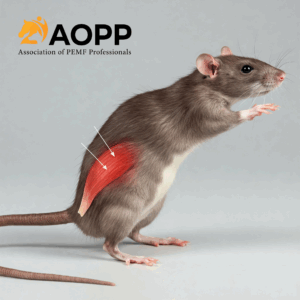
Before the surgical procedure, the animals were anesthetized with 80–100 mg/kg ketamine (Ketalar, Pfizer, Turkey) + 8–10 mg/kg xylazine (Xylazin Bio %2, Bioveta, Czech Republic) anesthesia and placed in the prone position with fixation of the tail and extremities. Skeletal muscle injury was created by using an 18 g biopsy needle on the right leg gastrocnemius muscle, approximately 3 mm proximal to the muscle-tendon junction of the biopsy needle. The lesion was made to cover roughly 50% of the cross-sectional area of the medial gastrocnemius muscle [14]. After the biopsy, the needle was removed, and an antiseptic dressing was applied. The wound was covered with a bandage for 2–3 days, and the bandaging was terminated after epithelialization was achieved and the bleeding stopped.
Half of the animals in the groups were sacrificed on the 7th day and the other half on the 14th day under the same anesthesia protocol. Following the abdominal incision, euthanasia was performed with blood taken from the inferior vena cava as a surgical exsanguination method. Half of the muscle tissues were placed in 10% formaldehyde for histopathological examination and sent to the relevant unit. The remaining half of the muscle tissues were put into − 20° for genetic analysis.
Stretching exercise protocol
In order to comfortably position the rats during the stretching exercise, 2 restraints were designed and produced with a 3D printer suitable for the body structure of the rats. Animals were comfortably and calmly placed in the restrainer before exercise. At the beginning of the application, soft tissue massage and mobilization were applied to the gastrocnemius muscle of the rats before the stretching exercise to prepare the tissue for exercise, and then gentle passive stretching was applied to the feet of the rats in the direction of 30° dorsi flexion [7]. The knee joint is extended during the stretching exercise. Stretching exercises were applied as 10 repetitions. Each repetition was performed as 1-minute of stretching and 30 s (s) rest. These processes took approximately 15 min for each rat [15, 16]. Stretching exercises were started the 3rd day after INJ in order to complete the inflammation process, strengthen the scar tissue and reduce the risk of recurrence [17]. Stretching practices were performed once a day, 5 days a week, and at the same times of the day. 5 sessions of stretching were applied to the rats sacrificed on the 7th day, and 10 sessions of stretching were applied to the rats sacrificed on the 14th day.Restrainers were cleaned daily with an antiseptic. All untrained rats were placed in the restrainer at the same time to be in the same conditions as the trained rats and to experience the same stress.
Discussion
The results of the study demonstrated that both stretching exercises and PEMF therapy resulted in a reduction in inflammation, degeneration, atrophy, and necrosis, while concurrently promoting accelerated muscle tissue recovery. In addition, by increasing the number of pycnotic nuclei, the muscle cell became more active, and the muscle mass increased. The application of PEMF and stretching exercise limited fibrosis formation by providing a controlled increase in VEGF and FGF, thus contributing to functional recovery. TGF-β1 level on both the 7th and 14th days; exercise, PEMF, and combined treatment groups were significantly reduced compared to the injury group, so single and combined treatment contributed to the reduction of fibrosis. It was observed that the single or combined use of stretching exercise and PEMF treatments significantly reduced eNOS levels compared to the injury group.
Inflammation is one of the first responses given at the tissue level when an injury occurs in any tissue due to various factors, such as trauma, as in skeletal muscle. In this inflammatory process, the body’s defense cells, especially neutrophils, migrate to the injured area and try to repair that area in order to minimize the damage. Depending on the severity and duration of the injury, these cells, which are collected at the scene, can deepen the inflammation during repair and accelerate regeneration by increasing the synthesis of growth factors such as VEGF and FGF. While this process may continue up to a certain level, if the irritation continues or due to the excessive severity of the injury, the intense inflammation may delay the repair. In order to ensure the restoration of the area, cytokines secreted from the defense cells coming to the scene can call other cells to the lesion area and increase the synthesis of growth factors [20]. In addition, in order to improve the migration to the lesion area during inflammation, NO is synthesized from the blood vessels via the eNOS enzyme, thereby expanding the vessel [21].
The presence of mononuclear cell infiltrations, atrophy, and necrosis detected in the INJ group induced by an 18 g biopsy needle in this study shows that the inflammatory muscle injury model required for stretching exercise and PEMF activities is formed. Both stretching exercise and PEMF applied in the treatment groups kept these inflammatory reactions at a lower level and accelerated muscle tissue recovery. In addition, by increasing the number of pyknotic nuclei, the muscle cell became more active, and the muscle mass increased. In order to increase the regeneration capacity of the injured muscle tissue, it is very important that these findings are parallel in both methods, and separate and intermittent applications of both methods can be recommended for the regression of inflammation. In addition, when combined treatment is applied, increasing the time between stretching exercise or applying stretching one day and PEMF the other day may facilitate tissue recovery and improve healing.
One of the main signs of cell death is nuclear changes that occur due to the fragmentation of chromatin and DNA. In pycnosis, which is also observed in the apoptotic process, the nucleus shrinks, and the chromatin density increases. The large pyknotic nuclei and high chromatin density in this study may also indicate that apoptosis, a negative effect for treatment, is reduced [22]. The fact that the number of pyknotic nuclei detected in this study increased in the single and combined treatment groups and was less in combined use than in single-use can be interpreted as suggesting single use for muscle development.
Being able to attenuate such an inflammatory response that can delay this repair time in the human body, which can repair itself, and reasonably reduce inflammatory cell infiltration is important to coordinate the inflammatory and regeneration process, which allows better functional recovery of the muscle after muscle injury [23]. In addition to routine stretching exercises, it is very important to support future studies to know that inflammation can be reduced more quickly and repair time can be accelerated with PEMF applications, which are easy to obtain and can be used repeatedly.
Berrueta et al. stated that stretching reduces inflammation, and active and passive stretching creates similar effects with a mechanical effect on tissues. In addition, it has been supported that stretching applied to the connective tissue reduces neutrophil migration within the tissue, which is a central aspect of the acute inflammatory response. Finally, stretching has been shown to have a direct mechanical effect on inflammatory regulatory molecules in connective tissue [24]. In other studies, both stretching exercises and exercises with a significant stretching component (e.g., yoga, Tai Chi) reduce circulating proinflammatory cytokine levels [25–27]. Achieving homeostasis in the face of acute inflammatory/immune challenges in the human body involves maintaining a balance of highly complex biochemical and cellular interactions, such as cytokine expression and signal transduction. The importance of maintaining healthy cytokine expression during this impactful time cannot be overstated. PEMF has the potential to regulate this very delicate balance [28]. In a rat rotator cuff injury and transosseous repair model to investigate the structural and functional effects of PEMF on tendon-to-bone healing, PEMF treatment was shown to improve recovery 3 months after surgery in patients with small or moderate rotator cuff tears [29]. In the study by Brown and Baker, calf muscle injury in rabbits was treated with shortwave diathermy (KDD) for 20 min twice a day for 8 and 16 days. After the treatment staining performed with the HE method and the results of the study indicated that 8-day treatment did not affect muscle recovery, the 16-day treatment program showed a trend towards increased recovery [30]. Contrary to the study of Brown and Baker [30], the decrease in the number of inflammatory cells detected on the 7th day in this study suggests that it may be related to the duration of PEMF use. The fact that different effects were observed at other times in the preliminary study also supports this result.
The increase in eNOS activity for repair at the site of inflammation in response to injury is a known physiological condition [31], and it was expected that PEMF application would deepen this increase [32–34]. However, the fact that the decrease in eNOS activities in PEMF groups was inversely related to the presence of inflammation within the scope of the study indicates that eNOS levels may decrease secondary to the anti-inflammatory activity of PEMF using other mechanisms. In many studies, PEMF, which increases eNOS increase-mediated repair upon healing, regressed inflammation in our study, resulting in a decrease in reactive endogenously synthesized eNOS levels. It is among the important outputs of the study that the stretching exercises act in parallel with the PEMF application and contribute positively to the repair process. In order to elucidate these situations, PEMF application times and different frequency values should be detailed with advanced molecular studies.
In the area of inflammation PEMF and stretching exercises limited the formation of fibrosis by providing a controlled increase in VEGF, which is an indicator of angiogenesis [35], and FGF, which performs fibroblast chemotaxis and is an indicator of fibrosis [20], thus contributing to functional recovery. The absence of an abnormal increase in the acute period and the fact that this increase becomes evident in the later period of the repair process can be interpreted as the regression of the inflammatory picture in the acute stage and the emphasis on the repair process in the later period.
In this study, VEGF levels in both INJ + EXE and INJ + PEMF groups and FGF levels in all treatment groups were higher than the control group, although not at a significant level, on the 7th day. On the 14th day, VEGF level increased in all groups except the control group while this increase was significantly higher in the injury group, except for the PEMF group, and FGF levels were significantly higher in the injury group than in all groups. As expected, the results support angiogenesis and wound healing, but the increase was more limited in combined treatments, contrary to expectations. We think that this may be due to the dosage of the treatment or the succession of applications without sufficient rest time. FGF level was also highest in the injury group on the 14th day. While the necessity of increasing growth factors has been shown in studies, it is not known exactly how much this increase should be and how long it should continue.
Several studies have shown that electrical stimulation of rat hind limb skeletal muscles can produce significant increases in the expression of the VEGF gene [36] and blood vessel density after stimulation [37, 38]. These results suggest that therapeutic modalities such as electrical stimulation of skeletal muscle may be promising to enhance angiogenesis following injury, potentially upregulating muscle regeneration events through stem cell activation and triggering of VEGF. In addition, mechanical stretching applied to muscle tissue can increase VEGF expression, while massage-like mechanical stretching can increase muscle regeneration through VEGF expression, stem cell, and inflammation modulation mechanism [39].
The amount of production of these factors may vary according to the type and severity of the injury, and it is known that intense VEGF and FGF increases also cause the development of fibrosis, which is an important problem in healing [20, 40]. Therefore, it is seen that the regular and certain level of expression of these growth factors rather than over or under production is more important. In this study, the application of both PEMF and stretching exercise provided a controlled increase in VEGF and FGF and limited the formation of fibrosis, thus giving the impression that it may have contributed to functional recovery.
Especially inadequacies in the repair process can cause the muscle tissue to heal with scarring and fibrosis. Different studies have been conducted to investigate the fibrosis formation process in muscle tissue and to determine the steps that cause it [41]. Current studies have focused on TGF-β1, which is an important mediator in the development of fibrosis, which is an undesirable condition in the healing of muscle injury. Many studies have shown that these protein’s levels increase in injury groups and cause fibrosis. Although various growth factors such as VEGF and FGF released from neutrophils, macrophages, fibroblasts, and myogenic precursors increase fibrosis, the most profibrogenic growth factor identified in the literature is TGF-β1 [42, 43].
Depending on the inflammatory milieu, injury can result either in a tissue’s complete regeneration or in its degeneration and fibrosis. TGF-β1 can modulate muscle repair not only by impacting at fibroblasts responsible for excessive extracellular matrix (ECM) deposition, but also by influencing the stem cells and inflammatory cells present and active within the injured area [44]. TGF-β1could prevent the apoptosis of Fibro-adipogenic progenitors (FAPs) and induce their differentiation into matrix-producing cells, which results in a shift from transient matrix deposition to persistent fibrogenesis [45].
It has been determined that TGF-β1 increases collagen synthesis in injured muscle tissue and plays an important role in the process leading to fibrosis, and antifibrotic agents have been used to inhibit this effect. However, studies have been conducted on the use of various biological agents to stimulate regeneration and contribute to the average satellite cell migration [12, 41, 46]. Stretching exercises, a cheaper treatment method, can be used for treatment as they can show positive effects at the cellular level during the acute recovery period. Although the benefits of stretching exercises on injured fiber are not fully known, a limited number of studies suggest that they can have healing-stimulating effects. The reason for this thought is that when stretching exercises are performed in accordance with the muscle architecture, they create parallel intermittent tension and, as a result, stimulate collagen synthesis. In addition, with passive stretching in the muscle, capillary density, fibril cross-sectional area, and glycogen content increase, resulting in hypertrophy [6]. When the literature is examined, it is seen that studies generally focus on anti-fibrotic agents, but there are limited studies on the effect of physiotherapy methods such as exercise and electro-physical agents on fibrosis. Some animal models have shown that passive movements result in a reduction of fibrosis in healing tendons [47]. In other studies evaluating the effect of active stretching exercises, it has been shown that subcutaneous collagen formation is reduced after injury [48, 49]. In a study using an ex vivo model, tissue stretching was shown to decrease soluble TGF-β1 [50]. Although more studies are needed, controlled stretching programs may be a promising treatment option for preventing and treating fibrosis [51]. Hwang et al. assessed the effectiveness of stretching and decorin application on fibrosis after laceration injury and stated that whenever stretching exercise was started, it reduced the level of fibrosis as much as decorin [7]. Techniques such as massage and massage-like mechanical stretching may decrease the duration of the fibrotic phase by increasing the circulating clearance of TGF-β [39]. There are some studies showing that PEMF can also change the TGF-β level in wounds. Although TGF-β is well known to promote fibroblast to myofibroblast differentiation, there is a lack of evidence that PEMF alters TGF-β levels in wounds [52]. Looking at other soft tissues, Jasti et al. [53]. stated that PEMF had no significant effect on the systemic expression of TGF-β in a rat tendinitis model. In this study, it was observed that TGF-β1 level on both the 7th and 14th days, there was a significant decrease in exercise, PEMF, and combined treatment groups compared to the injury group, so single and combined treatment contributed to the reduction of fibrosis. Our applications are non-invasive, affordable and easy to apply, and do not have any known side effects, showing their superiority compared to anti-fibrotic treatments. It is obvious that stretching exercises and/or PEMF application can provide therapeutic effects with anti-fibrotic effects in any disease that is likely to develop fibrosis.
The limitations of our study include the following: evaluation could not be made in the early stages of the inflammatory process; different evaluations were not able to determine the effect of oxidative stress on NO level; healing with treatments may have occurred in a shorter time than expected due to the small damage in the wound model; since the study was conducted on female rats, the estral cycle may affect the study model. For this reason, the study can also be conducted on male rats; and it is difficult to make accurate comparisons because different methods have been applied in evaluation and treatment in the literature. More research is required to establish the method’s effectiveness and examine various intracellular signaling pathways. The treatment protocols applied in this study were designed for animals. When the study will be carried out on humans, treatment protocols need to be rearranged.
Conclusions
This study showed that PEMF and stretching exercise may be effective in the treatment of muscle injuries, as they balance the inflammatory process in the muscle, have a positive effect on muscle development, accelerate healing, reduce TGF-β1 signaling, and prevent the development of fibrosis and inflammatory-induced eNOS activity. Both stretching exercises and PEMF can be used safely in the early period to accelerate the recovery and return to sports, which is the key point of rehabilitation, especially in professional athletes for whom even short periods of recovery are very important after muscle injuries. Further studies are needed to evaluate the effectiveness of these methods in more detail.
Mesut Ergan 1, Tahir Keskіn 2, İbrahim Aydın Candan 3, Yalçın Erzurumlu 4, Halil Aşci 5, Selçuk Çömlekçı 6, Ferdi Başkurt 7
PMID: 40128672 PMCID: PMC11931823 DOI: 10.1186/s12891-025-08442-0

